
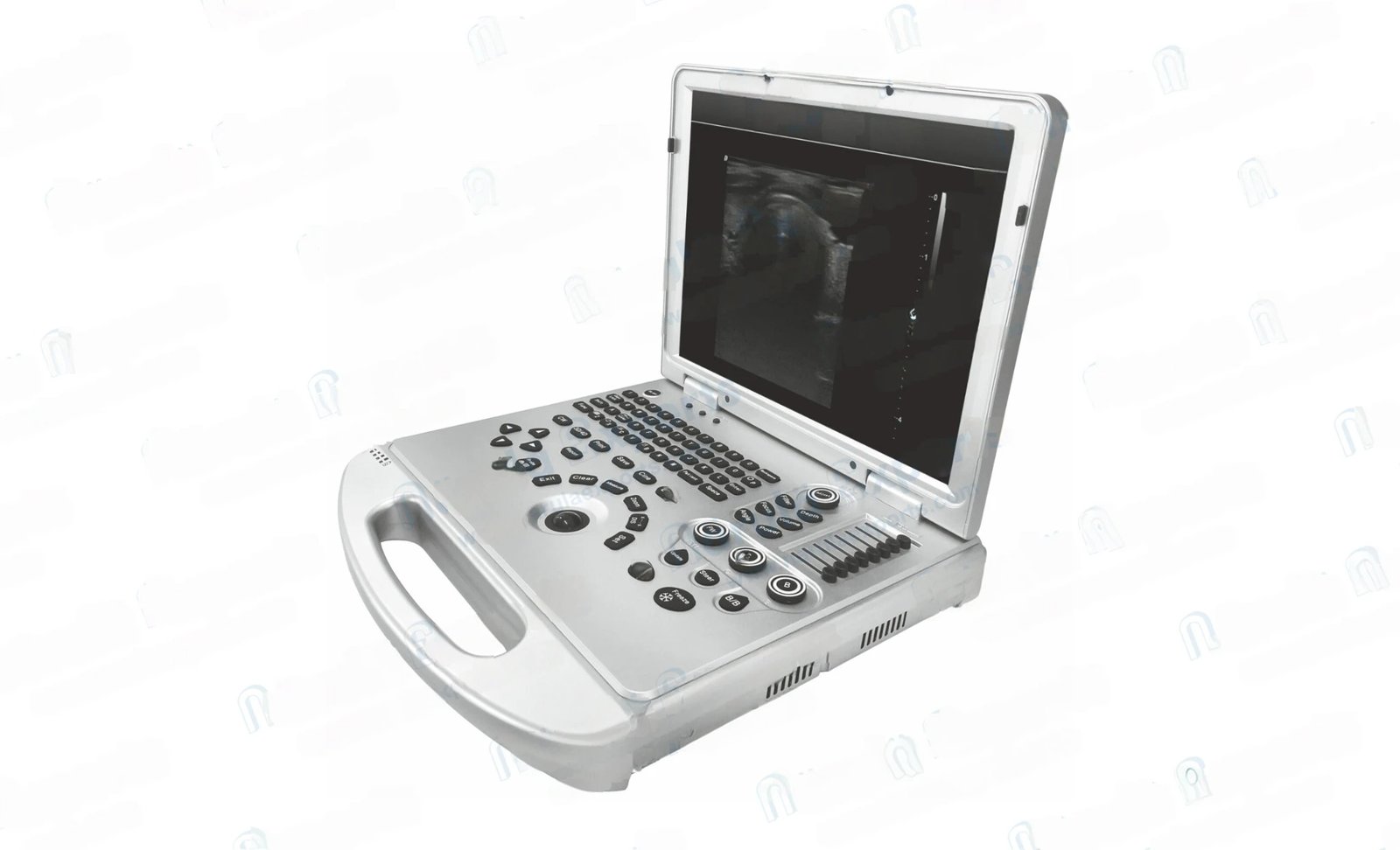
Model No.: 84-4090
The L3 has been designed from the relentless focus on delivering uncompromising performance at a cost-effective price Equipped with high-end imaging technology, color images more delicate, higher clarity. With ergonomic design, lightweight and compact, convenient for the use of medical staff in different scenarios; High resolution medical display, image loss free.
Smart Collaboration Reliable Operation
- 15 inch medical HD display
- One button image optimization
- TGC: details, customized image optimization
- Backlit control panel
- Large capacity data storage
- Trackball: for easy operation
Smooth Workflow
One-click intelligent optimization, fast access to quality images all-in-one clipboard Smooth processing Edge enhancement processing The host built-in SSD≥128G is fast and stable to start Cine playback: ≥4000 frames
Excellent Image Quality
Spectral pulse Doppler Directional energy Doppler Spatial composite imaging Tissue harmonic imaging technique 4B imaging mode
User-Friendly Operation
Backlit, easy-to-use control panel the classical ergonomic design With DICOM3.0 protocol, PACS system can be connected
Clear Image Visualization
The research and development has spent three years, integrating the most advanced design concept and technological innovation, to create full digital high performance full digital color Doppler ultrasound diagnostic instrument.
Intelligent operation process, humanized appearance design and intimate human-computer interaction as a whole, so that doctors in the process of clinical diagnosis will focus on the patient itself.

Micron Imaging Technology
Micro imaging technology, real-time tracking of different tissue edge specific signals, to achieve edge enhancement, while monitoring every pixel; The internal signals of the organization are optimized and the edge information and the internal pixel information are perfectly fused to restore the real and delicate 2D image with excellent hierarchical contrast.
Tissue harmonic imaging (THI)
By improving tissue contrast resolution, spatial resolution and eliminating near-field artifact, image clarity can be improved. It is mainly used in the diagnosis of cardiovascular and abdominal diseases, and plays an important role in the evaluation of lesion areas and demarcation of difficult imaging. This technology has been fully recognized by clinicians.
Harmonic technology retains the second harmonic signal to the maximum extent on the basis of removing the fundamental signal, which increases the signal intensity by more than 30% compared with the traditional signal processing, reduces noise and artifacts, and improves the contrast resolution of tissue image.

Trapezoid Imaging
It is a kind of extended imaging. On the basis of the original rectangle, it is transformed into trapezoid. The left and right sides are expanded to a certain extent to achieve a wider visul field.
The principle of ultrasound imaging is to use ultrasonic beam scanning organs, through the reception and processing of reflected signals, to obtain images of internal organs.

Carotid Spectrum
Spectral ultrasonography of carotid artery can provide a noninvasive, simple and reproducible method for the diagnosis of atherosclerosis. However, multi-parameter analysis should be advocated in the analysis of detection results. Besides the flow velocity of relevant vascular segments, pusing index, spectral morphology, blood flow direction and blood flow sound should also be considered.
Carotid ultrasound is helpful to determine the nature of the ischemic cerebrovascular disease of carotid artery atheromatous plaque and stability, and to determine the degree of carotid atherosclerosis and carotid stenosis, especially in the display has the advantages on the change of the arterial wall structure, for the early prevention and treatment of atherosclerosis provide objective basis, actively treating atherosclerosis and carotid stenosis in preventing ischemic brain have important significance.

HD Liver Imaging Effect
2D real-time ultrasound imaging is mainly used for the change of liver morphology. Ultrasound examination shows the pathological image of liver, which belongs to the change of acoustic physical properties. For the same lesion, different stages of disease development, Ultrasonic image performance is different.


Imaging with clarity and accuracy
Transducers

Across a wide range of clinical specialties: To meet the basic hospital in the abdomen, obstetrics, gynecology, urinary system, small organs, superficial, vascular, pediatric, newborn, muscle, physical examination and other aspects of the examination and diagnosis.